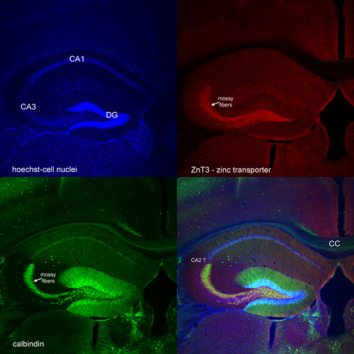
It’s fun to zoom out and get the big picture sometimes. This is one such picture I took long ago when I wanted to see if staining for zinc transporter 3 effectively labels the mossy fiber axons of the dentate gyrus. You can see by the perfect overlap with calbindin that it does the job, though the staining wasn’t quite as bright and obvious as calbindin. The abundance of zinc in mossy fiber axons is one of the peculiarities of the DG and it underlies numerous synaptic properties of DG neurons.
I think the goal was to build on previous work by Lipp, Ramirez-Amaya, and Routtenberg showing that spatial learning causes “sprouting” of mossy fibers, though when I found out that this phenomenon does not occur in mice the project was aborted.
But what else can you see in this picture?
- clear differential expression of calbindin: DG (lots) > CA1 > CA3 (none), and a scattering of strongly-positive interneurons (e.g. 5 cells where CA3 and CA1 meet)
- in CA1 you can see calbindin is expressed only in the lower band of cells (see hi res photo if needed; there is a ref for this, somewhere)
- a thin band of calbindin-positive fibers crossing the corpus callosum (CC)
- A small group of cells that are not contacted by the calbindin-positive mossy fiber axons (i.e. beyond CA3) yet do not express somatic calbindin (as seen in CA1). I’m guessing this may be mysterious and ambiguous field CA2.
Great pictures, Jason. I really like the “10,000 foot view”.
Pingback: Sunday Links #2 « Neuromancy Blog
Pingback: Thoughts on the mind, 6/28 « Children of Cajal