Now that I have something to show for it, let this be a formal announcement that I’ve returned to Toronto to join Paul Frankland’s lab (and therefore the larger Josselyn-Frankland group). I’ve always liked their work and one of the techniques I’m excited to learn is the use of viruses to alter gene expression in neurons. BECAUSE THIS WILL ALLOW ME TO TAKE PRETTY PICTURES!!! I will also say that this will be a short (but hopefully sweet) stay as I’ll be leaving at the end of the year to start my own lab in the Psychology Department at the University of British Columbia (!).
Now, on with the pictures! As always, I recommend high-res viewing (click on the image to view it, bigger, on Flickr).
Using a retrovirus, which infects dividing cells, I made the amazing discovery of four adult-born cells which all had the exact same shape and were located right next to each other!
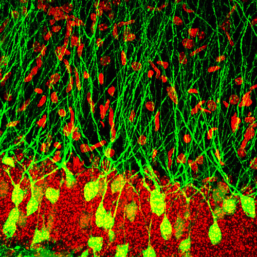
More dentate granule cells, infected with Herpes Simplex Virus and expressing GFP.
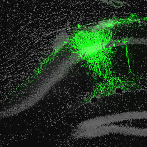
In the process of learning the surgeries required to stereotactically inject the virus, you inevitably target the wrong regions. Which is fun because then you get a glimpse of something new. Here are GFP+ CA1 pyramidal neurons. You can see their axons projecting down and to the left, towards the subiculum.
More CA1 pyramidal neurons from the same animal but an adjacent section. I love the fanning of the dendrites. Click on the image and you can see spines in a higher resolution version.
Every day I see a retrovirally-labelled, non-neuronal (?) cell that looks more beautiful than the last one I imaged. Makes me want to cry.
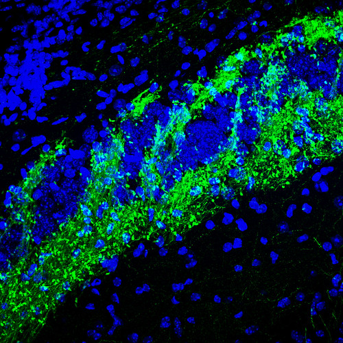
 Here are dentate granule cell axons, the mossy fibers, flowing through CA3 pyramidal neuron cell bodies, like a river gushing over rocks. In the woods. The balls on a string appearance is caused by the infamously large presynaptic boutons, which are distributed along the axon (like balls on a string, thin rope, or even dental floss).


Congrats on the lab at UBC, Jason! You earned it. I’m sure that you will do some great science there.
Congrats on the job!! BC is so gorgeous and I hear it’s a fun place to live too. Plus: your own lab! Super exciting.
Also, I aspire to take pictures half as pretty as yours. Unfortunately, my near future is filled with light instead of confocal microscopy.
Awesome pictures, Jason! And congrats on the new gig! I expect a seminar invite to UBC to be forthcoming…
Which will of course come in the form of a Twitter @ reply
Wow! Amazing work, I am a UofT undergraduate in the Behaviour Genetics and Neurobiology Program. Your work is such an inspiration and this website puts a whole new spin on science! Thank you for your inspirational work !
Thank you Aya! It is also a huge inspiration to hear from you! I’m glad you enjoy it.
that is quite amazing to see 4 near identical newborn GCs. did you inject a cocktail of 4 retroviruses?
No! I just had some fun with Photoshop!
Hi Jason
I am a Doctoral student at Experimental Neurlogy, University hospital, Jena Germany working on the same topic …functional neurogenesis . I am always following your work and your web page… its really splendid. I also aspire to do some good neuroscience and you for sure inspire me for doing that…
Best!!
Varunika – thank you for your encouraging words! I’m glad you enjoy the blog and best of luck with your research!
Hello, Jason
In stumbling across your pic of four “newborn” adult neurons of identical shape, I am reminded of an experiment from Frank Solomon’s lab (MIT) in the ’70s (Cell 16:165;1979) where he observed that neuroblastoma cells pass the memory of their dendritic morphology to daughter cells after mitosis. Although these cells as a population are highly diverse in shape, upon initial emergence from mitosis, sibling cells can adopt the same complex shape that can be identical or mirror image. Perhaps the simultaneous transition to a postmitotic state of sibling neural precursors freezes the morphological signature.
Very interesting blog. I will starting checking in more frequently.
Mitch Goldfarb