Most studies of adult neurogenesis are concerned with neuronal age. Or at least they should be. This is because new neurons develop from a stage where they have no excitatory synapses to one where they have many. If we assume the traditional view that information is stored at excitatory synaptic connections, then young neurons are initially useless and only become physiologically and behaviorally meaningful when they have matured to a point where they can relay and process information. It is therefore critical that the developmental timecourse of new neurons be mapped out, so we know when new neurons become functionally relevant, or whether they might even have different functions at different ages.
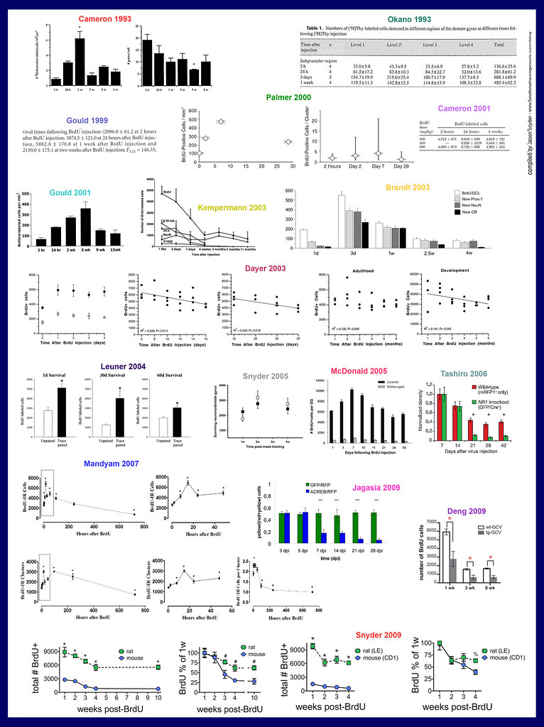
Below are what I hope to be comprehensive visual collages of all published timecourse experiments, where a certain property of new neurons is examined at multiple (≥ 3) different ages. They are grouped by studies of: 1) cell survival, 2) marker expression, 3) functionality, and 4) miscellaneous studies that do not quite fit into the first 3 categories. I’ve ordered the data roughly chronologically and have included the first author’s name and publication year so you can read deeper, if needed. Indeed, if you know these studies already, a brief look at the graphs will bring back the take home message. However, since the data is stripped of text, if the studies are unfamiliar, you’ll have to go to the original source to figure out what the heck they mean (use Pubmed to at least obtain abstracts for the original studies if I didn’t provide a direct link).
Personally, I like timecourse studies for the same reason I like to have all my music albums or books visible at the same time: at a single glance they provide a lot of information – each individual stage of maturation can be interpreted within a bigger picture. The result of these many hours of work will either be a) that the purpose of adult neurogenesis will become immediately clear, or b) that we’ll all have some fancy collages to pin on our bulletin boards and look intelligent.
The survival timecourse
New neurons are born and then many die. The survival timecourse answers the questions: How many new neurons are born? Where are they born and where do they end up, anatomically? How many of them survive and can their survival be altered? Survival timecourses are typically performed by injecting animals with a mitotic marker that will label new neurons as they’re being born, e.g. ³H-thymidine (old school), BrdU (tried and true – example), or a GFP-expressing retrovirus (new school). At a later date one can then detect these birthdated new neurons and count them, see where they’re located etc.
What do these survival timecourses tell us?
- many newborn neurons die between 1w and 4w of age but after that they all survive
- neurons born during infancy are an exception as they DO die off many months after their birth (Dayer 2003), lending support to the sexy-but-underexplored idea that neuronal turnover might underlie memory turnover in the hippocampus
- the number of new cells labeled with a birthdating marker (e.g. BrdU) grows between 2 hours and several days after the birthdating marker is administered
- this is caused by continued division of the stem cell or precursor cell that took up the marker in the first place (see expression timecourse, below). After a few cell divisions the marker gets diluted to undetectable levels.
- the general timecourse of cell death is similar in young and aged animals (McDonald 2005) and in mice and rats, although more cells die in mice (Snyder 2009)
- the addition and culling of newborn neurons in the monkey hippocampus (Gould 2001) follows a delayed timecourse compared to rodents
- CREB signalling is critical for neurons to survive between 5-7 days old (Jagasia 2009), NMDA receptors are critical for survival from 14-21 days (Tashiro 2006)
- thus, CREB signalling would appear to regulate survival before new neurons have formed excitatory connections and are functional (see below) and NMDA receptors regulate survival during the early phase of excitatory synapse formation, when new neurons are just beginning to be able to contribute to behavior. Knowing how to regulate neuronal survival has obvious implications for disorders where reduced neurogenesis might be a causative factor.
- learning increases the survival of new neurons (Leuner 2004)
- learning does not increase the survival of new neurons (Snyder 2005)
The expression timecourse
All cell types within the body express different genes/proteins that serve the cell’s function. Since a muscle cell has a completely different function than a skin cell, it will naturally express different proteins. In like manner, a 1 week-old neuron is functionally distinct from a 4 week-old neuron and the two will also express different proteins (to some extent). Many people have taken advantage of this, using these different proteins as markers that identify a new cell as a neuron vs. a glial cell or, more specifically, an immature neuron vs. a mature neuron. By simultaneously visualizing (via immunohistochemistry) both the birthdating marker (e.g. BrdU) and these phenotypic markers, one can know both the exact age of the neuron and its general degree of maturity. For a 10 sec guide to cell labeling with BrdU and phenotypic markers, see here.
What do these expression timecourses tell us?
- some markers (proteins) are increasingly expressed as new neurons mature over 4 weeks (NSE, NeuN, calbindin)
- other markers are mainly expressed when new neurons are < 4 weeks-old (DCX, PSA-NCAM, calretinin)
- most studies have used the same markers (e.g. DCX, NeuN) to simply demonstrate that new cells are neurons, but some have examined expression of markers that are associated with a more specific function, such as glucocorticoid receptors (Cameron 1993, Garcia 2004) or vascular markers (Palmer 2000)
- BrdU (or other birthdating markers) labeled cells express cell division markers (e.g. Ki67) several days after BrdU is administered. This does not mean newborn neurons are dividing – what it represents is the continued division of the stem cell, or precursor cell, that was originally labelled. (therefore you can never know the exact age of a new cell, but pretty close)
The functional timecourse
The previous timecourses are all well and good but sheer numbers of cells say nothing about whether the new neurons actually work. And markers like DCX or NeuN may give a general hint at the maturity of a neuron but not much more. Functional timecourses address these gaps. A direct measure of function would be whether a new neuron has electrophysiological properties that enable it to process information (e.g. input and output synapses, action potentials). Less direct signs of function can be inferred from the morphology of a new neuron and whether a new neuron is capable of expressing activity-dependent immediate early genes.
What do these functional timecourses tell us?
- electrophysiology (Esposito 2005; Ge 2006), anatomy/morphology (Hastings 1999; Zhao 2006; Toni 2007; Toni 2008), and activity-dependent gene expression (Jessberger 2003; Snyder 2009) all point to new neurons forming their first synapses at 2-4 weeks of age
- around 4 weeks of age, new neurons go through a phase where they have enhanced synaptic plasticity (Ge et al 2007) and enhanced activation during behavior (Snyder 2009)
- thus, at a young age, new neurons are more modifiable by experience than mature neurons. This may enable them to make a greater impact on behavior, either at this age or by shaping their further integration into the circuitry so they can alter brain function when fully mature and functional
- young neurons have distinct neurotransmitter profiles: initially they receive GABAergic inputs and later receive excitatory glutamatergic inputs (Esposito 2005). Notably, GABA depolarizes immature neurons (Ge 2006), unlike its typically-inhibitory effects on mature neurons. Also, immature neurons have a unique form of the NMDA receptor (NR2B), which endows them with their enhanced plasticity (Ge 2007).
- blocking CREB signalling (Jagasia 2009) or the depolarizing effects of GABA (Ge 2006) inhibits the functional maturation of new neurons
- by 8 weeks of age, new neurons are pretty much fully developed, though Zhao 2006, Toni 2007 and Toni 2008 show that 8-10 week-old neurons are still slightly underdeveloped, presynaptically and postsynaptically
- does this mean that 8-10 week-old neurons, despite no longer having enhanced synaptic plasticity or enhanced activation during behavior, might still function differently than fully mature neurons?
Other timecourses
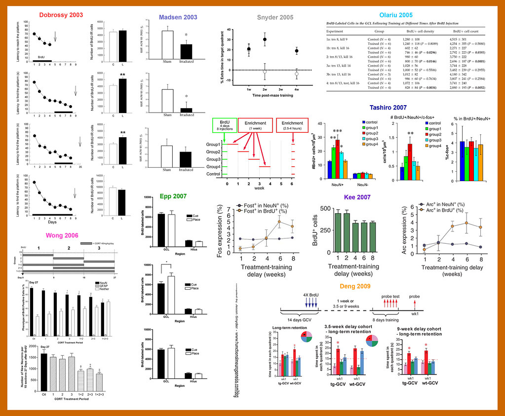
There are some timecourse-ish studies that, instead of examining new neurons of different ages, have examined the final fate of same-aged neurons that had been manipulated at different stages of their development. From the first 3 figures we can see that specific stages during a new neuron’s development are associated with enhanced plasticity, unique neurotransmitter profiles and increased likelihood of cell death. Therefore, it is very possible that the ultimate fate of an adult-born neuron depends on when experiences occur, relative to these different stages.
What do these timecourses tell us?
Several show us that experience can modify the number of new neurons, but that the magnitude and direction of the change depends on how old the neurons are when the animal undergoes the experience. For example:
- environmental enrichment enhances survival of new neurons mainly when the neurons are 1-2 weeks old (Tashiro 2007)
- spatial learning in the water maze enhances survival when the neurons are 5-10 days old (Epp 2007) or 1-2 weeks old (Kee 2007)
- the stress hormone corticosterone decreases new neuron survival when administered for 18-day, but not 9-day, stints during new neuron development (Wong 2006)
- Dobrossy 2003 and Olariu 2005 show the interesting but difficult-to-interpret findings that neuron addition can be increased or decreased depending on the extent of learning and the age of the neuron relative to the learning experience
- The Kee 2007 data suggests that fully mature, 10-week-old neurons are activated during memory retrieval only if they were old enough, at the time of learning, to be involved in forming the original memory
———————————————————————
Hopefully it’s now clear that a 3 day-old neuron differs from a 3-week-old neuron from a 3-month-old neuron. We have seen examples where the developmental stage of a new neuron influences it’s functional maturation and survival, clues that could someday be used to manipulate adult neurogenesis for therapeutic purposes.
Lastly, comparing many of these timecourses side by side, I’m reminded why I like them so much: I trust them. By examining the same thing at multiple time points, each study inherently has a lot of controls. If you take a single time point out of some of these studies, say the % of new neurons that have excitatory glutamatergic inputs at 14 days-old in Esposito 2005 and Ge 2006, you might wonder, who’s right here? One finds 5%, the other finds 70%. I am curious as to why they differ but I still trust both of these studies and refer to them often because, within their respective timecourses, both 14-day data points make sense and, across studies, the 2 timecourses themselves do generally agree, even if they are slightly shifted.
Update April 12, 2016: we have followed up on this story by examining the survival timecourse for neurons born at the peak of postnatal dentate gyrus development. The short version is that it’s a different story in development.


I wish I had read this post when I was still in the lab.
Check out my (the company’s) blog at http://www.cerescanimaging.com/Cerescan-Blogs.aspx
Seems like you are doing well and educating mightily as usual. Keep up the good posts and I look froward to the future ones.
Woah, very sweet. The “brdu + doublecortin + zif268” picture is especially useful. And you’re right about the issue of trust… time course studies seem to be particularly well controlled.
Pingback: The first example of functional neurogenesis? | Functional Neurogenesis
We just couldnt leave your website before saying that I really enjoyed the quality information you offer to your visitors… Will be back often to check up on new stuff you post!
Very informative!
did this take you awhile? ha
Yes it took a long time. A very long time. Did it take you a long time to leave that comment?
Yes. Also, it seems as if you could make a review paper out of this
Thank you so much for taking the time to gather all this data and put it together in a such an informative way. It is one thing to read a paper or two, but when you see the whole picture put together like this it is amazing! This piece of work is highly commendable and very exciting to read. Thanks again, and please continue this superb work.
Ha! Thanks a lot! Those were fun days, having the time to compile the essentials of such an insane number of studies. They probably need updating by this point!
Pingback: Nice Enhance The Brain photos | Cognitive Enhancer
Thanks for this superinformative piece. It’s really helpful.
Quick questions: What genes would you suggest being studied for both hippocampal and Olfactory bulb neurogenesis if all you have is regular PCR or qPCR?
Also, can one reliably say that SVZ/Olfactory bulb neurogenesis reflect/correlates with hippocampal neurogenesis?
Kindly reply here and to the email supplied.
Thank you.
Pingback: mεdıan voıd sεmıotıcs . . thε “sεlƒmε” | Best blog on memory efficiency
Very helpful, It’s a really good work.
thank you!
Pingback: Survival of neurons born in development vs. adulthood