![]() I recently became re-acquainted with the neurogenesis literature while writing the last post, re-finding data in papers whose gist, but not details, I had remembered. I reached out a little bit, asking others if I had forgot any studies and indeed I had, including this study by Okano, Pfaff and Gibbs from 1993.
I recently became re-acquainted with the neurogenesis literature while writing the last post, re-finding data in papers whose gist, but not details, I had remembered. I reached out a little bit, asking others if I had forgot any studies and indeed I had, including this study by Okano, Pfaff and Gibbs from 1993.
I’ve been interested in new neuron function since 1999 and so I’m actually quite surprised I missed this study until so recently. In 1999 the neurogenesis literature was so scant that it was easy to know ALL of the studies, even the early Altman, Kaplan and Nottebohm studies from the 1960s through 1980s. Even studies that were not interesting were interesting, because there was nothing else to read! So, had I known about it back then, I would have been pretty interested in this study by Okano et al. if only for its focus on cell cycle markers. But I really would have been interested in it because it has a small functional experiment that was way ahead of it’s time:

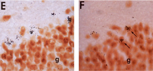
(Figure Legend) E, Thymidine-labeled/Fos-negative cells detected in the subgranular region 3 d after receiving ³H-Thy and 3 hr after pentylenetetrazolinduced seizure activity. Note the presence of many Fos-positive cells in the adjacent granule cell layer. F, Thymidine-labeled/Fos-positive cells detected in the granule cell layer 4 weeks after receiving ³H-Thy and 3 hr after pentylenetetrazol-induced seizure activity. g, granule cell layer.
(Results) To determine if the newly differentiated neurons were functional, we examined the expression of Fos-IR in response to pentylenetetrazole-induced seizure activity. In the absence of induced seizures, very few (< 1%) ³H-Thy-labeled cells in the dentate gyrus were immunoreactive for Fos-IR at any of the six time points examined. However, within 3 hr following seizure activity, an induction of Fos-IR within 21 .O% (at 1 week post-thymidine injection) and 81.3% (at 4 weeks post-thymidine injection) of the ³H-Thy-labeled cells detected in the granule cell layer was observed (Fig. 3E,F), suggesting that these cells had formed functional connections.
Fos is one of a number of immediate-early genes (IEGs) that are expressed following synaptic activity. IEGs allow short-term changes in synaptic function to turn into long-term changes (which is thought to be required for short-term memory to transform into long-term memory). IEGs can therefore be used to identify neurons that are contributing to memory formation. Okano et al. noted that < 1% of new cells expressed Fos when rats were not stimulated overtly. It could have been that the rats’ experience prior to death was not memorable or it could have reflected the fact that only a fraction of hippocampal neurons are activated by normal experiences. To get around this they activated all neurons that were possibly “activateable”, by using a convulsant. Therefore, all neurons that had synapses expressed Fos and a timecourse of neuronal maturation, one that fits nicely into the subsequent literature, was obtained. When I say subsequent literature I’m referring to examples like the next study which used IEGs to characterize new neuron maturation, 10 years later. And also the many electrophysiological studies that emerged beginning 10-15 years later. It’s funny that none of them referred to this study. Had I known about it I certainly would have cited it as potential evidence that new neurons in rats mature faster than in mice (21% of 7-day-old rat neurons expressed Fos – higher than I’ve observed here but consistent with a recent rat electrophysiology study?)
Now, this is a small experiment but how did it go unnoticed when so many people are curious about the function of adult neurogenesis? Well, the study does focus mainly on the expression of cell cycle markers and less on neuronal function – most of the 102 subsequent papers that cite it are cell cycle studies. But, a number of familiar papers do cite it – papers I thought I’d read thoroughly back when there was nothing else to read on the subject, so it’s just proof you can never totally be on top of things…hence the often-used “To the best of our knowledge, this is the first example of….”
Oh, and this is not the first example of functional neurogenesis. That title goes to Paton & Nottebohm who, to the best of my knowledge, demonstrated it for the first time in songbirds.
Reference:
Okano HJ, Pfaff DW, & Gibbs RB (1993). RB and Cdc2 expression in brain: correlations with 3H-thymidine incorporation and neurogenesis. The Journal of neuroscience : the official journal of the Society for Neuroscience, 13 (7), 2930-8 PMID: 8331381
To my knowledge, Imayoshi et al (2008) were the first to provide evidence of what newborn adult mammalian neurons actually do. But then, your knowledge of this field is almost certainly way more comprehensive than mine, so I might be wrong.
Yeah, I would say Imayoshi et al. is one study out of many that have attempted to figure this out!